Why Are Cytotoxic Carbon NanoTubes or NanoWorms Found In mRNA Vaccines?
Updated: Aug 29, 2021
Published: 2021, Apr 13
Author: Robert O Young CPC, MSc, DSc, PhD, Naturopathic Practitioner
www.drrberyoung.com
https://rumble.com/vg0gul-what-is-a-carbon-nanotube-or-nano-worm-doing-in-a-vaccine.html
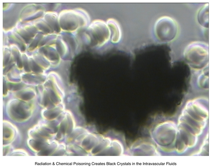
Dr. Robert Young, please identify the black nano worms in the Pfizer vaccine as seen under pHase contrast microscopy?

In the phase contrast microscopy video above, you have asked me to review and comment.
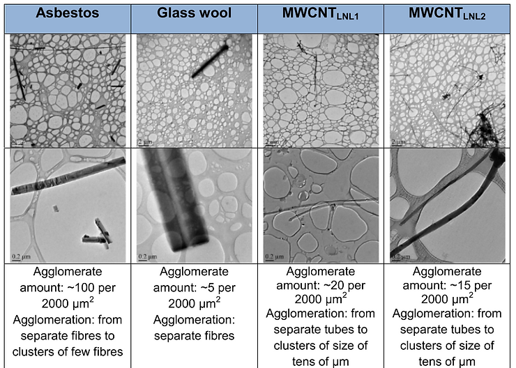
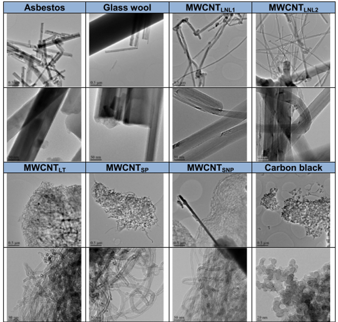
I would suggest the following: what you are viewing is a carbon multi-walled nanotube (MWCNTs) (graphene oxide – GO) which I have shown to elicit asbestos-like toxicological effects on cell membranes and cell genetics.

To reduce the risk for humans and animals, I would suggest that the physicochemical characteristics or reactivity of nano materials should be used to predict a serious health hazard.
Fibre-shape and the ability to generate reactive oxygen species (ROS) are important indicators of highly acidic hazardous materials. Asbestos is one of those known toxic acidic ROS generators, while MWCNTs may either produce or scavenge ROS.
However, certain biomolecules, such as albumin – used as dispersants in nano material or particulate preparations for toxicological testing in vivo and in vitro – may reduce the surface reactivity of these nano materials.
Testing MWCNT materials induced highly variable cytotoxic effects which generally are related to the abundance and characteristics of agglomerates/aggregates and to the rate of sedimentation.
I have found that All carbon nano materials – MWCNTs, like the ones many people are asking about in the above video, will scavenge hydroxyl radicals which is a major alkaline buffer (OH-) released by lymphocytes to protect the alkaline design of the body fluids, especially the Interstitial fluids of the Intersitium.
I would suggest that this is for the purpose of reducing proton/hydrogen concentrations in the body fluids, leading to the risk of decompensated acidosis in all of the fluids or solutions of the body which I have tested, including the vascular and interstitial fluids of the Interstitium, leading to pathological blood coagulation, hypoxia and death by suffocation in humans and animals.
The effect of bovine serum albumin (BSA) in cell culture medium with and without BEAS 2B cells on radical formation/scavenging by five MWCNTs, Printex 90 carbon black, crocidolite asbestos, and glass wool, using electron spin resonance (ESR) spectroscopy, showed cytotoxic effects measured by trypan blue exclusion assay among the materials tested. Two types of long, needle-like MWCNTs (average diameter > 74 and 64.2 nm, average length 5.7 and 4.0 μm, respectively) induced, in addition to a scavenging effect, a dose-dependent formation of a unique, yet unidentified radical or antioxidant release in both absence and presence of cells, which also coincides with cytotoxicity of these nanotubes or in simple terms MWCNTs are a contributing factor in the cause of a pathological blood coagulation, hypercapnia, hypoxia, and death by suffocation. These are all the symtoms of CoV – 2 now called CoV – 19.

Based upon the microscopic evaluation presented in the above video it is my professional opinion that what is being viewed is a MWCNTs or a carbon nanotube, like graphene oxide, which is highly cytotoxic or an acidifier to blood, interstitial fluid compartments and intracellular fluids, which may lead to cell membrane degeneration and genetic mutation of the body cells putting at risk the healthy state of all glands, organs and tissues in humans and animals.

Watch, Listen, Learn, Care and Share with everyone you love and care about! – www.drrobertyoung.com
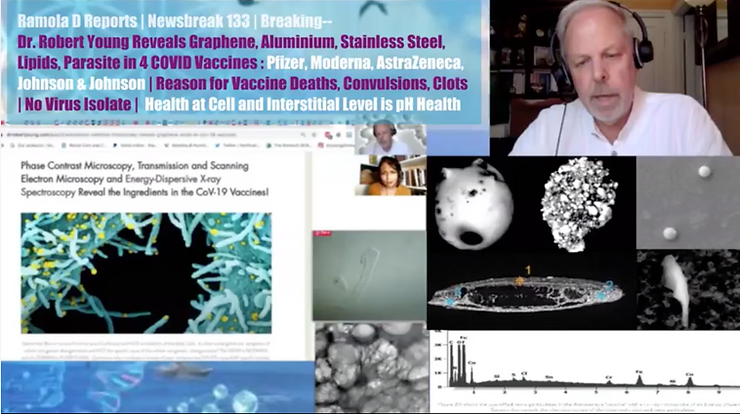
SCIENCE TEAM REVEALS GRAPHENE, ALUMINUM, LNP CAPSIDS, PEG & PARASITES IN 4 CoV – 19 VACCINES
Watch, Listen and Learn the Truth About Four Big Pharma CoV – 19 Vaccines by Clicking on the Picture Below:

https://www.bitchute.com/video/Z2sAH0Woz38r/ Absolutely a Bombshell!
Major revelations on what is in the CoV – 2 – 19 vaccines, with the use of electron, pHase, dark field, bright field and other types of microscopy from the original research of Dr. Robert Young and his scientific team, confirming what the La Quinta Columna researchers found – toxic nanometallic content with magneticotoxic, cytotoxic and genotoxic effects, as well as identified life-threatening parasites.
A Life Changing Life Saving Revelation This is major scientific revelation: please watch, listen, read and learn and then please share this video and article far and wide!

A Life Changing Life Saving Revelation This is major scientific revelation: please watch, listen, read and learn and then please share this video and article far and wide!
LINKS FOR MORE VIDEOS AND ARTICLES:

https://www.bitchute.com/video/jqD5tbQO1WUT/
Dr. Young’S Scientific Teams major article reporting their findings: Click below to read the entire article: https://www.drrobertyoung.com/post/transmission-electron-microscopy-reveals-graphene-oxide-in-cov-19-vaccines Dr. Young’s Scientific Teams article revealing graphene and other non-disclosed ingredients in a PDF format:
References
1. De Volder MFL, Tawfick SH, Baughman RH, Hart AJ: Carbon nanotubes: present and future commercial applications. Science 2013, 339:535–539.
2. Liu Y, Zhao Y, Sun B, Chen C: Understanding the toxicity of carbon nanotubes. Acc Chem Res 2012, 46:702–713.
3. Fenoglio I, Aldieri E, Gazzano E, Cesano F, Colonna M, Scarano D, Mazzucco G, Attanasio A, Yakoub Y, Lison D, Fubini B: Thickness of multiwalled carbon nanotubes affects their lung toxicity. Chem Res Toxicol 2011, 25:74–82.
4. Service RF: Nanotubes: the next asbestos? Science 1998, 281:941.
5. Lam C-W, James JT, McCluskey R, Hunter RL: Pulmonary toxicity of singlewall carbon nanotubes in mice 7 and 90 days after intratracheal instillation. Toxicol Sci 2004, 77:126–134.
6. Shvedova A, Castranova V, Kisin E, Schwegler-Berry D, Murray A, Gandelsman V, Maynard A, Baron P: Exposure to carbon nanotube material: assessment of nanotube cytotoxicity using human keratinocyte cells. J Toxicol Environ Health A 2003, 66:1909–1926.
7. Kim J, Song K, Lee J, Choi Y, Bang I, Kang C, Yu I: Toxicogenomic comparison of multi-wall carbon nanotubes (MWCNTs) and asbestos. Arch Toxicol 2012, 86:553–562.
8. Palomäki J, Välimäki E, Sund J, Vippola M, Clausen PA, Jensen KA, Savolainen K, Matikainen S, Alenius H: Long, needle-like carbon nanotubes and asbestos activate the NLRP3 inflammasome through a similar mechanism. ACS Nano 2011, 5:6861–6870.
9. Poland CA, Duffin R, Kinloch I, Maynard A, Wallace WAH, Seaton A, Stone V, Brown S, MacNee W, Donaldson K: Carbon nanotubes introduced into the abdominal cavity of mice show asbestos-like pathogenicity in a pilot study. Nat Nano 2008, 3:423–428.
10. Sakamoto Y, Nakae D, Fukumori N, Tayama K, Maekawa A, Imai K, Hirose A, Nishimura T, Ohashi N, Ogata A: Induction of mesothelioma by a single intrascrotal administration of multi-wall carbon nanotube in intact male Fischer 344 rats. J Toxicol Sci 2009, 34:65–76.
11. Takagi A, Hirose A, Futakuchi M, Tsuda H, Kanno J: Dose-dependent mesothelioma induction by intraperitoneal administration of multi-wall carbon nanotubes in p53 heterozygous mice. Cancer Sci 2012, 103:1440–1444.
12. Takagi A, Hirose A, Nishimura T, Fukumori N, Ogata A, Ohashi N, Kitajima S, Kanno J: Induction of mesothelioma in p53+/- mouse by intraperitoneal application of multi-wall carbon nanotube. J Toxicol Sci 2008, 33:105–116.
13. Xu J, Futakuchi M, Shimizu H, Alexander DB, Yanagihara K, Fukamachi K, Suzui M, Kanno J, Hirose A, Ogata A, et al: Multi-walled carbon nanotubes translocate into the pleural cavity and induce visceral mesothelial proliferation in rats. Cancer Sci 2012, 103:2045–2050.
14. Murphy FA, Poland CA, Duffin R, Al-Jamal KT, Ali-Boucetta H, Nunes A, Byrne F, Prina-Mello A, Volkov Y, Li S, et al: Length-dependent retention of carbon nanotubes in the pleural space of mice initiates sustained inflammation and progressive fibrosis on the parietal pleura. Am J Pathol 2011, 178:2587–2600.
15. Murphy F, Poland C, Duffin R, Donaldson K: Length-dependent pleural inflammation and parietal pleural responses after deposition of carbon nanotubes in the pulmonary airspaces of mice. Nanotoxicology 2012, 1:11.
16. Nagai H, Okazaki Y, Chew SH, Misawa N, Yamashita Y, Akatsuka S, Ishihara T, Yamashita K, Yoshikawa Y, Yasui H, et al: Diameter and rigidity of multiwalled carbon nanotubes are critical factors in mesothelial injury and carcinogenesis. Proc Natl Acad Sci 2011, 108:E1330–E1338.
17. Nagai H, Toyokuni S: Differences and similarities between carbon nanotubes and asbestos fibers during mesothelial carcinogenesis: Shedding light on fiber entry mechanism. Cancer Sci 2012, 103:1378–1390.
18. Sargent L, Reynolds S, Castranova V: Potential pulmonary effects of engineered carbon nanotubes: in vitro genotoxic effects. Nanotoxicology 2010, 4:396–408.
19. Shvedova AA, Pietroiusti A, Fadeel B, Kagan VE: Mechanisms of carbon nanotube-induced toxicity: Focus on oxidative stress. Toxicol Appl Pharmacol 2012, 261:121–133.
20. Sund J, Alenius H, Vippola M, Savolainen K, Puustinen A: Proteomic characterization of engineered nanomaterial–protein interactions in relation to surface reactivity. ACS Nano 2011, 5:4300–4309.
21. Jaurand M-C, Renier A, Daubriac J: Mesothelioma: Do asbestos and carbon nanotubes pose the same health risk? Part Fibre Toxicol 2009, 6:1–14.
22. Kamp DW, Graceffa P, Pryor WA, Weitzman SA: The role of free radicals in asbestos-induced diseases. Free Radic Biol Med 1992, 12:293–315.
23. Fenoglio I, Greco G, Tomatis M, Muller J, Raymundo-Piñero E, Béguin F, Fonseca A, Nagy JB, Lison D, Fubini B: Structural defects play a major role in the acute lung toxicity of multiwall carbon nanotubes: physicochemical aspects. Chem Res Toxicol 2008, 21:1690–1697.
24. Jacobsen NR, Pojana G, White P, Møller P, Cohn CA, Smith Korsholm K, Vogel U, Marcomini A, Loft S, Wallin H: Genotoxicity, cytotoxicity, and reactive oxygen species induced by single-walled carbon nanotubes and C60 fullerenes in the FE1-Muta™Mouse lung epithelial cells. Environ Mol Mutagen 2008, 49:476–487.
25. Bihari P, Vippola M, Schultes S, Praetner M, Khandoga A, Reichel C, Coester C, Tuomi T, Rehberg M, Krombach F: Optimized dispersion of nanoparticles for biological in vitro and in vivo studies. Part Fibre Toxicol 2008, 5:1–14.
26. Elgrabli D, Abella-Gallart S, Aguerre-Chariol O, Robidel F, Rogerieux F, Boczkowski J, Lacroix G: Effect of BSA on carbon nanotube dispersion for in vivo and in vitro studies. Nanotoxicology 2007, 1:266–278.
27. Vippola M, Falck G, Lindberg H, Suhonen S, Vanhala E, Norppa H, Savolainen K, Tossavainen A, Tuomi T: Preparation of nanoparticle dispersions for invitro toxicity testing. Hum Exp Toxicol 2009, 28:377–385.
28. NANOGENOTOX: Facilitating the safety evaluation of manufactured nanomaterials by characterizing their potential genotoxic hazard. Nancy: Bialec; 2013.
29. Foucaud L, Wilson MR, Brown DM, Stone V: Measurement of reactive species production by nanoparticles prepared in biologically relevant media. Toxicol Lett 2007, 174:1–9.
30. International Agency for Research on Cancer: Man-made vitreous fibres. Lyon: International Agency for Research on Cancer; 2002 [ IARC Monographs on the Evaluation of Carcinogenic Risks to Humans, vol 81.].
31. Jacobsen NR, White PA, Gingerich J, Møller P, Saber AT, Douglas GR, Vogel U, Wallin H: Mutation spectrum in FE1-MUTATMMouse lung epithelial cells exposed to nanoparticulate carbon black. Environ Mol Mutagen 2011, 52:331–337.
32. Ellinger-Ziegelbauer H, Pauluhn J: Pulmonary toxicity of multi-walled carbon nanotubes (Baytubes®) relative to α-quartz following a single 6 h inhalation exposure of rats and a 3 months post-exposure period. Toxicology 2009, 266:16–29.
33. Jensen KA: Summary report on primary physicochemical properties of manufactured nanomaterials used in NANOGENOTOX. NANOGENOTOX Final Report 2013: [http://www.nanogenotox.eu/files/PDF/Deliverables/ d4.1_summary%20report.pdf].
34. Murray A, Kisin E, Tkach A, Yanamala N, Mercer R, Young S-H, Fadeel B, Kagan V, Shvedova A: Factoring-in agglomeration of carbon nanotubes and nanofibers for better prediction of their toxicity versus asbestos. Part Fibre Toxicol 2012, 9:10.
35. Searl A, Buchanan D, Cullen RT, Jones AD, Miller BG, Soutar CA: Biopersistence and durability of nine mineral fibre types in rat lungs over 12 months. Ann Occup Hyg 1999, 43:143–153.
36. Saber A, Jensen K, Jacobsen N, Birkedal R, Mikkelsen L, Møller P, Loft S, Wallin H, Vogel U: Inflammatory and genotoxic effects of nanoparticles designed for inclusion in paints and lacquers. Nanotoxicology 2012, 6:453–471.
37. Roche M, Rondeau P, Singh NR, Tarnus E, Bourdon E: The antioxidant properties of serum albumin. FEBS Lett 2008, 582:1783–1787.
38. Pacurari M, Yin X, Zhao J, Ding M, Leonard S, Schwegler-Berry D, Ducatman B, Sbarra D, Hoover M, Castranova V, Vallyathan V: Raw single-wall carbon nanotubes induce oxidative stress and activate MAPKs, AP-1, NF-kappaB, and Akt in normal and malignant human mesothelial cells. Environ Health Perspect 2008, 116:1211–1217.
39. Bennett SW, Adeleye A, Ji Z, Keller AA: Stability, metal leaching, photoactivity and toxicity in freshwater systems of commercial single wall carbon nanotubes. Water Res 2013, 47:4074–4085.
40. Carella E, Ghiazza M, Alfè M, Gazzano E, Ghigo D, Gargiulo V, Ciajolo A, Fubini B, Fenoglio I: Graphenic nanoparticles from combustion sources scavenge hydroxyl radicals depending upon their structure. Bio Nano Sciences 2013, 3:112–122.
41. Kagan VE, Tyurina YY, Tyurin VA, Konduru NV, Potapovich AI, Osipov AN, Kisin ER, Schwegler-Berry D, Mercer R, Castranova V, Shvedova AA: Direct and indirect effects of single walled carbon nanotubes on RAW 264.7 macrophages: Role of iron. Toxicol Lett 2006, 165:88–100.
42. Mercer R, Hubbs A, Scabilloni J, Wang L, Battelli L, Schwegler-Berry D, Castranova V, Porter D: Distribution and persistence of pleural penetrations by multi-walled carbon nanotubes. Part Fibre Toxicol 2010, 7:28.
43. Henstridge MC, Shao L, Wildgoose GG, Compton RG, Tobias G, Green MLH: The electrocatalytic properties of Arc-MWCNTs and Associated ‘Carbon Onions’. Electroanalysis 2008, 20:498–506.
44. Ambrosi A, Pumera M: Amorphous Carbon Impurities Play an Active Role in Redox Processes of Carbon Nanotubes. J Phys Chem C 2011, 115:25281–25284.
45. He H-y, Pan B-c: Studies on structural defects in carbon nanotubes. Front Phys China 2009, 4:297–306.
46. van Berlo D, Clift M, Albrecht C, Schins R: Carbon nanotubes: an insight into the mechanisms of their potential genotoxicity. Swiss Med Wkly 2012, 142:w13698.
47. Tsuruoka S, Takeuchi K, Koyama K, Noguchi T, Endo M, Tristan F, Terrones M, Matsumoto H, Saito N, Usui Y, et al: ROS evaluation for a series of CNTs and their derivatives using an ESR method with DMPO. J Phys: Conference Series 2013, 429:012029.
48. Srivastava R, Pant A, Kashyap M, Kumar V, Lohani M, Jonas L, Rahman Q: Multi-walled carbon nanotubes induce oxidative stress and apoptosis in human lung cancer cell line-A549. Nanotoxicology 2010, 5:195–207.
49. Reddy ARN, Reddy YN, Krishna DR, Himabindu V: Multi wall carbon nanotubes induce oxidative stress and cytotoxicity in human embryonic kidney (HEK293) cells. Toxicology 2010, 272:11–16.
50. Lindberg HK, Falck GCM, Singh R, Suhonen S, Järventaus H, Vanhala E, Catalán J, Farmer PB, Savolainen KM, Norppa H: Genotoxicity of short single-wall and multi-wall carbon nanotubes in human bronchial epithelial and mesothelial cells in vitro. Toxicology 2013, 313:24–37.
51. Miserocchi G: Physiology and pathophysiology of pleural fluid turnover. European Respiratory Journal 1997, 10:219–225.
52. Porter D, Hubbs A, Chen B, McKinney W, Mercer R, Wolfarth M, Battelli L, Wu N, Sriram K, Leonard S, et al: Acute pulmonary dose–responses to inhaled multi-walled carbon nanotubes. Nanotoxicology 2012, 7:1179–1194.
53. Shen J-W, Wu T, Wang Q, Kang Y: Induced stepwise conformational change of human serum albumin on carbon nanotube surfaces. Biomaterials 2008, 29:3847–3855.
54. Yang M, Meng J, Mao X, Yang Y, Cheng X, Yuan H, Wang C, Xu H: Carbon Nanotubes Induce Secondary Structure Changes of Bovine Albumin in Aqueous Phase. Journal of Nanoscience and Nanotechnology 2010, 10:7550–7553.
55. Salvador-Morales C, Townsend P, Flahaut E, Vénien-Bryan C, Vlandas A, Green MLH, Sim RB: Binding of pulmonary surfactant proteins to carbon nanotubes; potential for damage to lung immune defense mechanisms. Carbon 2007, 45:607–617.
56. Wang F, Yu L, Monopoli MP, Sandin P, Mahon E, Salvati A, Dawson KA: The biomolecular corona is retained during nanoparticle uptake and protects the cells from the damage induced by cationic nanoparticles until degraded in the lysosomes. Nanomedicine: Nanotechnology, Biology and Medicine 2013, 9:1159–1168.
57. Reddel RR, Ke Y, Gerwin BI, McMenamin MG, Lechner JF, Su RT, Brash DE, Park JB, Rhim JS, Harris CC: Transformation of human bronchial epithelial cells by infection with SV40 or adenovirus-12 SV40 hybrid virus, or transfection via strontium phosphate coprecipitation with a plasmid containing SV40 early region genes. Cancer Res 1988, 48:1904–1909.
58. Park MVDZ, Neigh AM, Vermeulen JP, de la Fonteyne LJJ, Verharen HW, Briedé JJ, van Loveren H, de Jong WH: The effect of particle size on the cytotoxicity, inflammation, developmental toxicity and genotoxicity of silver nanoparticles. Biomaterials 2011, 32:9810–9817. doi:10.1186/1743-8977-11-4
59. Nymark et al.: Free radical scavenging and formation by multi-walled carbon nanotubes in cell free conditions and in human bronchial epithelial cells. Particle and Fibre Toxicology 2014 11:4.


