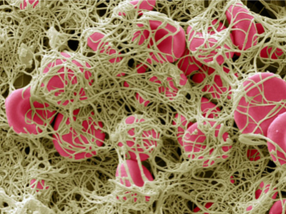
The Significance of the D-dimer Blood Test in Covid -19 Dis-ease
Updated: Feb 11, 2023
Oct 21, 2021

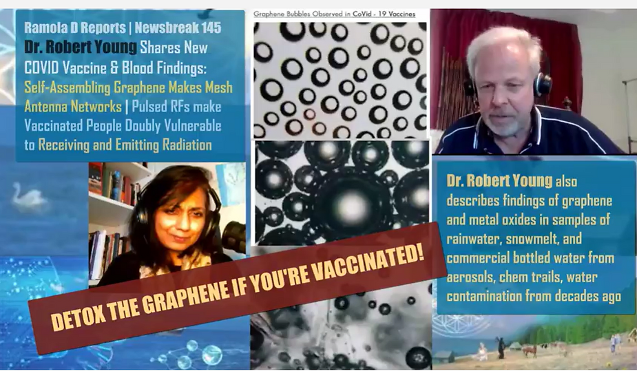
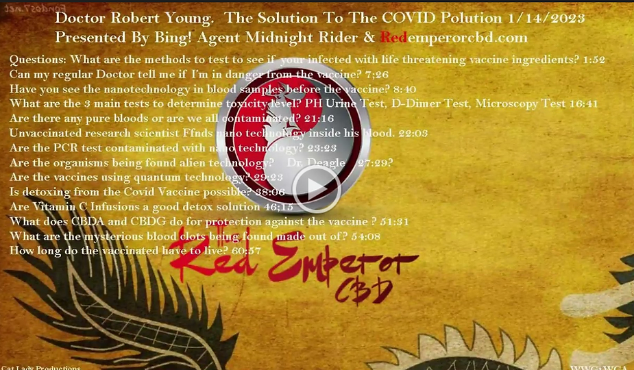
In this podcast, we talk with Dr. Robert Young who explains the significance of the D-dimer blood test as it relates to Covid-19 disease, pathological coagulation and blood clots leading to hypoxia and death. Click on the link below or any of the diagrams to listen and learn more about the D-dimer blood test and pathological blood clotting.
Click hear to listen: https://freedomfromaddiction.libsyn.com/
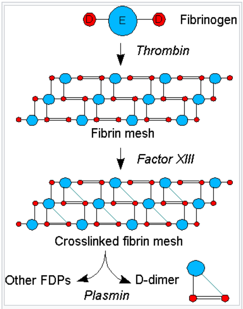
Dr. Young is a world-class scientific investigator and expert in pathological blood coagulation especially on microscopic and submicroscopic elements of the human blood.

He has written and published hundreds of scientific papers. You can find out more about Dr. Robert O. Young at www.drrobertyoung.com/blog.

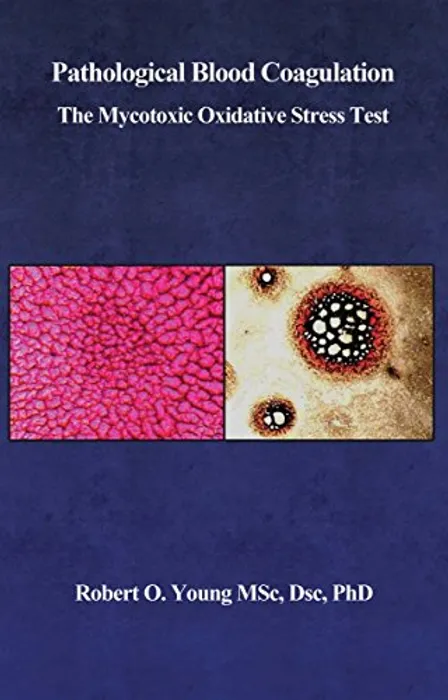
Check out Dr. Robert O. Young’s peer-reviewed article below on “pathological blood coagulation” and a revolution way of viewing the process of healthy or unhealthy coagulation of blood viewed under brightfield microscopy with low magnification. [8]
Micro and Nanographs of Graphene Nanowires, Threads, Ribbons, Dots and Bubbles of the Vaccinated Observed in LIVE CAPPILARY BLOOD BLOOD CLOTS UNDER Brightfield Microscopy at Low Magnification!
Graphene Based Nanowires are superconductor batteries used for tissue scaffolding inside the human body and for receiving and transmitting data across the ‘Internet of Things’.
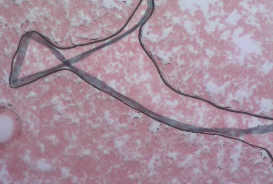
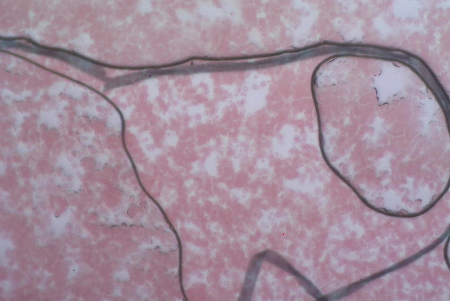
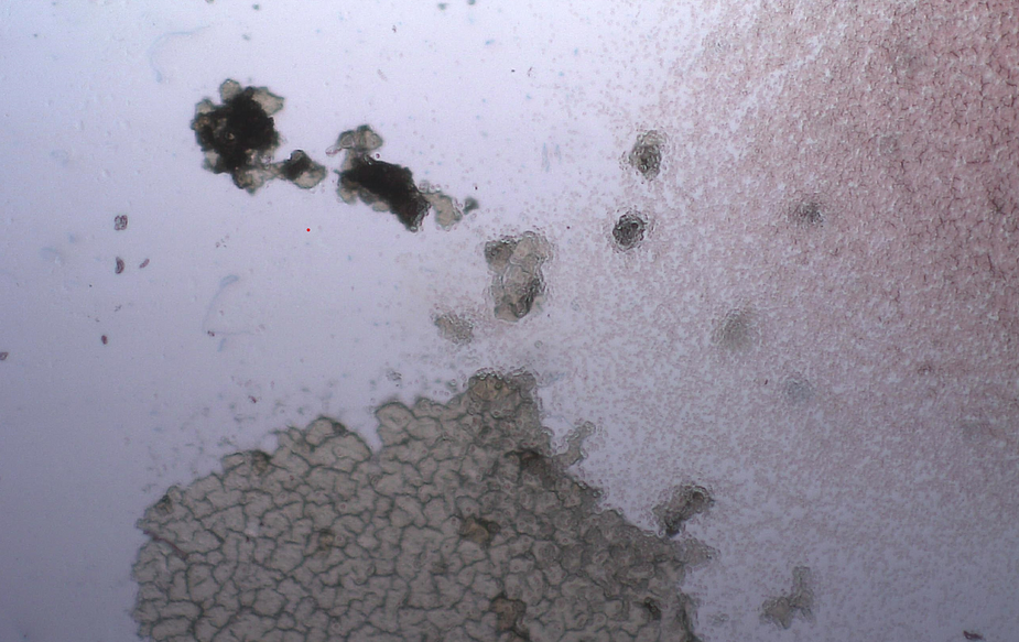
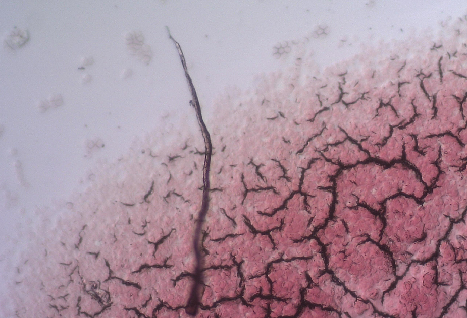
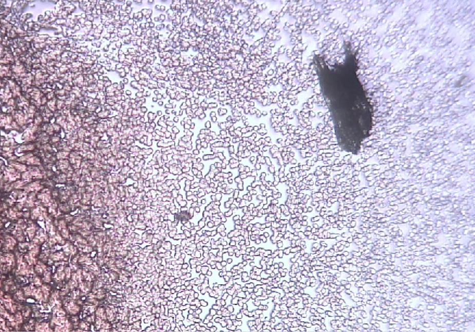
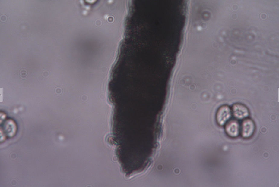
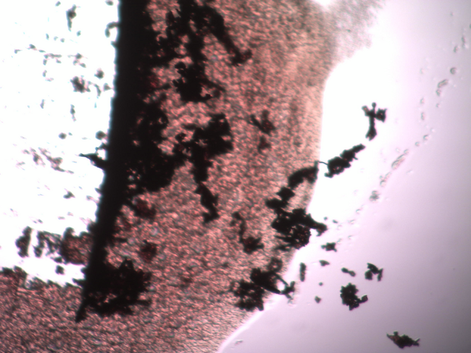
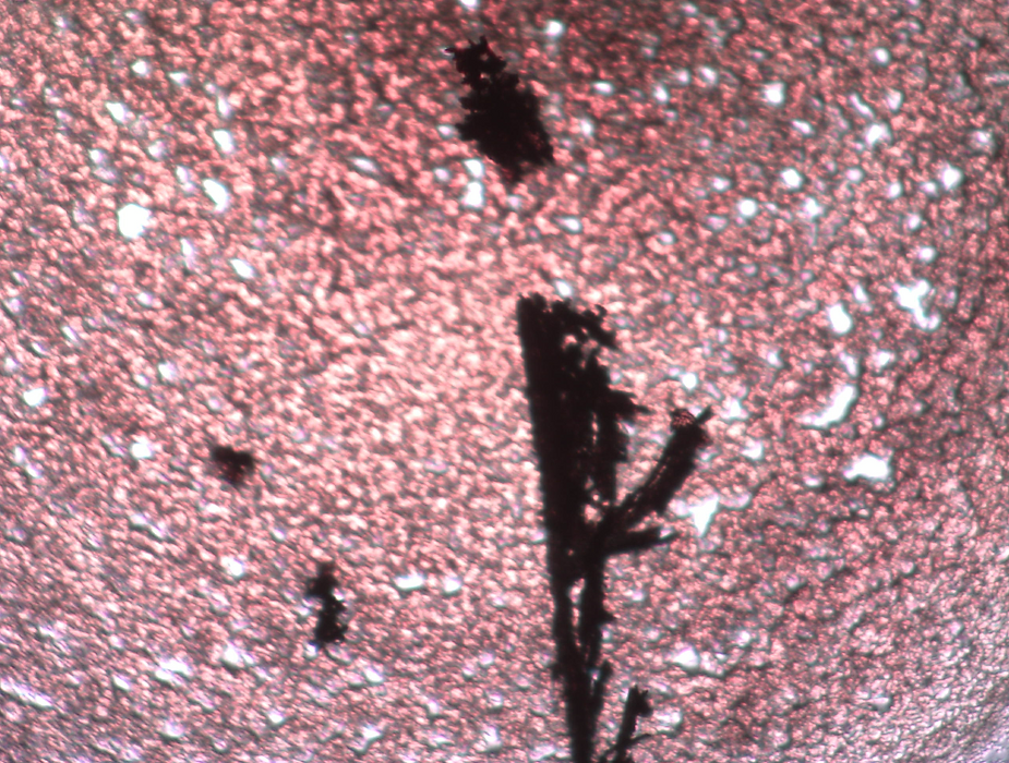

GRAPHENE NANOWIRES, RIBBONS AND NANO NETWORKS IN THE BLOOD COMBINES WITH EMF 5G RADIATION CAUSES OF INJURY AND DEATH!

Different morphology of nano graphene oxide as wires, ribbons, tubes, dots and sheets as observed in the live unchanged and unstained capillary blood of a VAXXinated female.
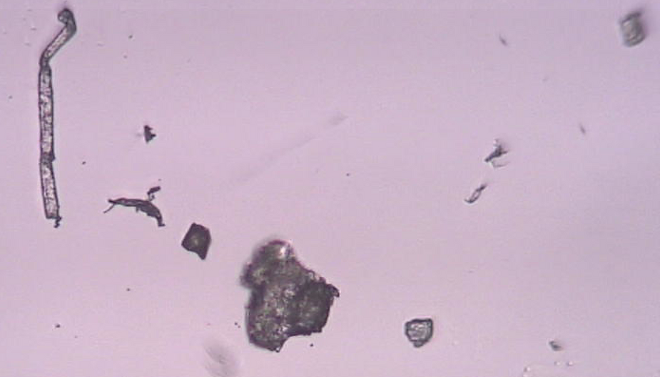
Nano Graphene Oxide Nanowires and Threads as observed in the live unchanged and unstained capillary blood under pHase contrast microscopy from a VAXXinated female.
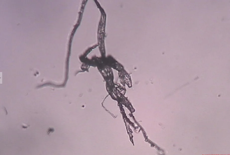
Nano Graphene Oxide sheets, tubes, nanowires, threads and dots observed in the live unchanged capillary blood under pHase contrast microscopy from a VAXXinated female. Please note the red blood cell clots throughout the smear.
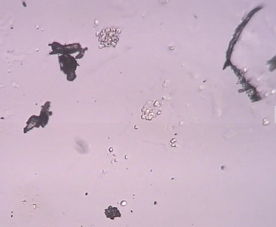
A large Nano Graphene Oxide Tubular blood clot observed in the live unchanged and unstained capillary blood under pHase contrast microscopy from a VAXXinated male.
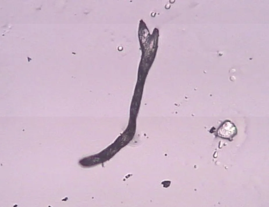
A large fibrous graphene oxide micro blood clot observed in the live unchanged and unstained capillary blood of a VAXXinated female under pHase contrast microscopy.
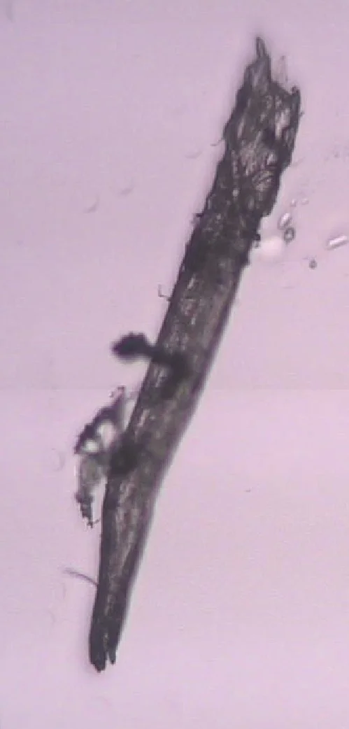
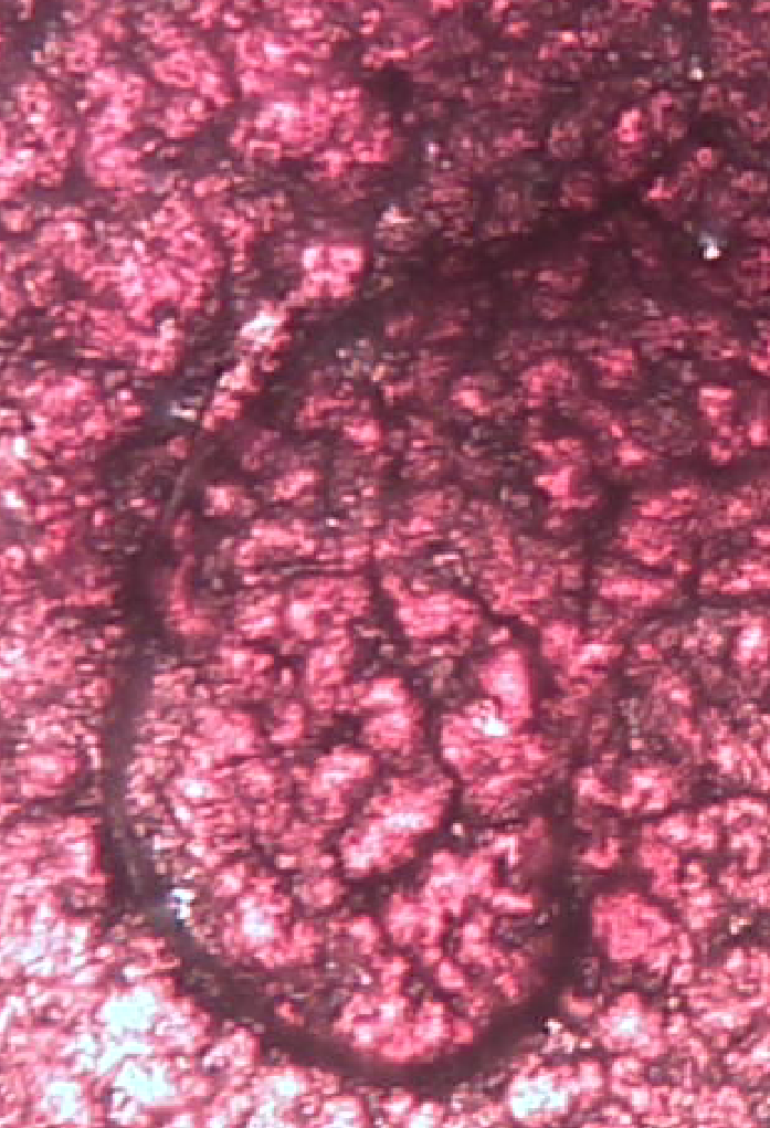
A pathological capillary blood clot containing graphene oxide threads and tubes observed in the live unchanged and unstained blood of a VAXXinated female under brightfield microscopy.
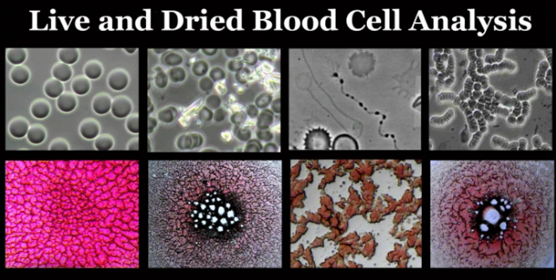
Micrographs of Healthy and UnHealthy Blood Under pHase Contrast and Brightfield Microscopy
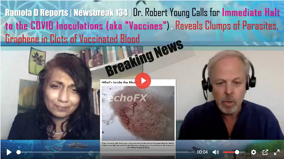
Graphene in Nano and Micro Blood Clots Viewed in the VAXXinated and UNVAXXinated Capillary Blood!
In the Following Video Dr. Young Shares His Work, Research and Findings in a Demonstration of Live and Dried Blood Analysis Using Phase Contrast and Brightfield Microscopy:
Learn More About the Work, Research and Findings of Dr. Robert O. Young

Robert O Young MSc, DSc, PhD, Naturopathic Practitioner www.drrobertyoung.com

Follow Dr. Robert O. Young on Twitter at: https://twitter.com/phmiraclelife
You can support the research of Dr. Robert O. Young with your prayers and donations at: https://www.givesendgo.com/research

https://rumble.com/v18ku7h-5g-radiation-poisoning-combined-with-graphene-poisoning-of-the-blood.html
Understand Why Blood Clots Form Inside the Blood Vessels!
Read Dr. Robert O. Young’s Peered Review Scientific Research Article Published in the International Journal of Vaccines and Vaccination on Pathological Blood Coagulation! (2016)
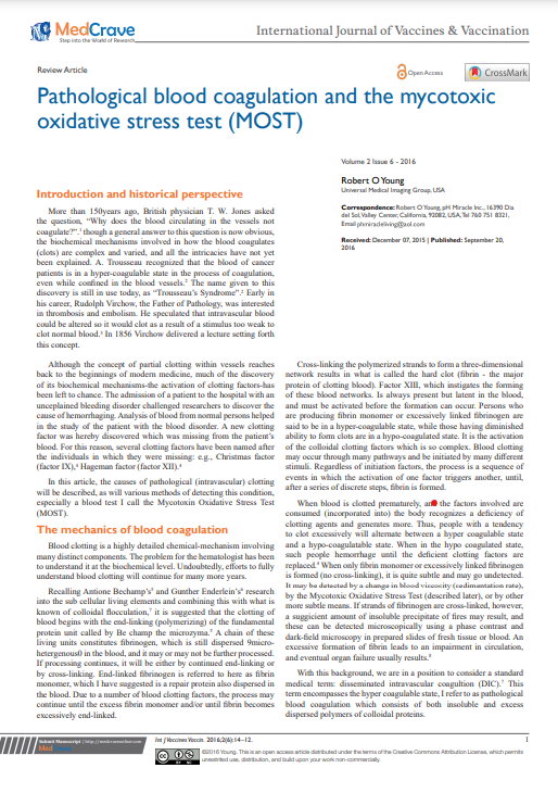
Pathological Blood Coagulation and the Mycotoxic Oxidative Stress Testing, Young RO (2016) Pathological Blood Coagulation and the Mycotoxic Oxidative Stress Test (MOST). Int J Vaccines Vaccin 2(6): 00048. DOI: 10.15406/ijvv.2016.02.00048
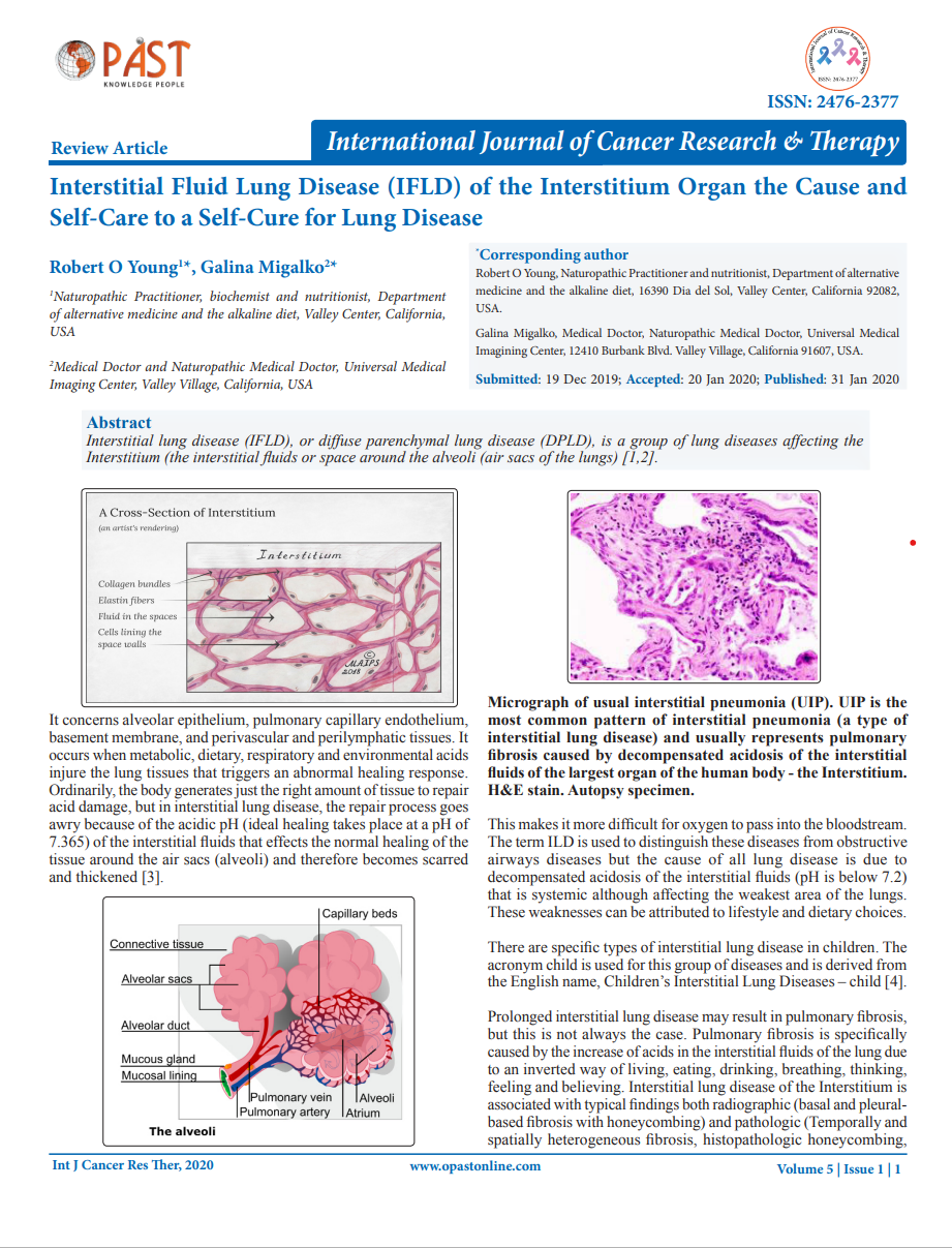
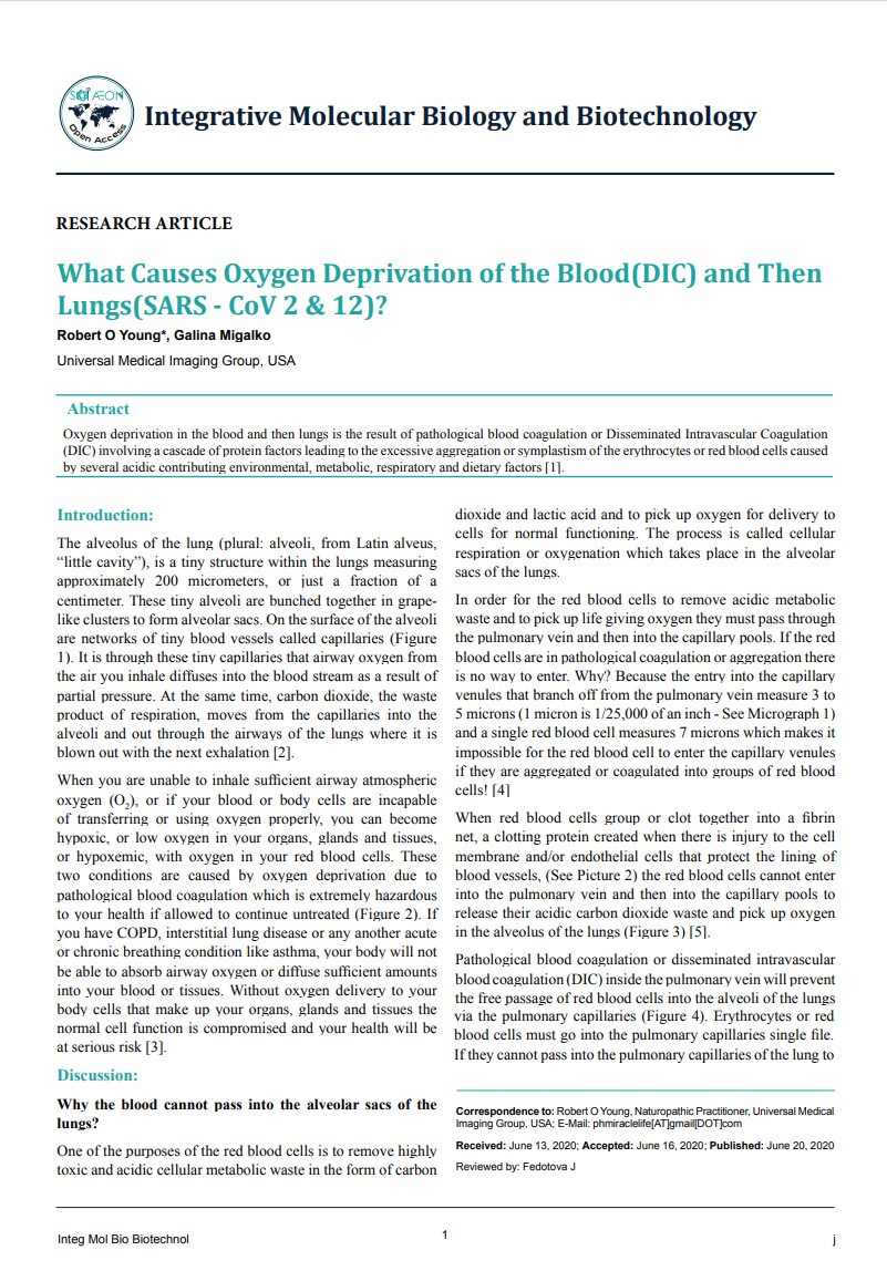
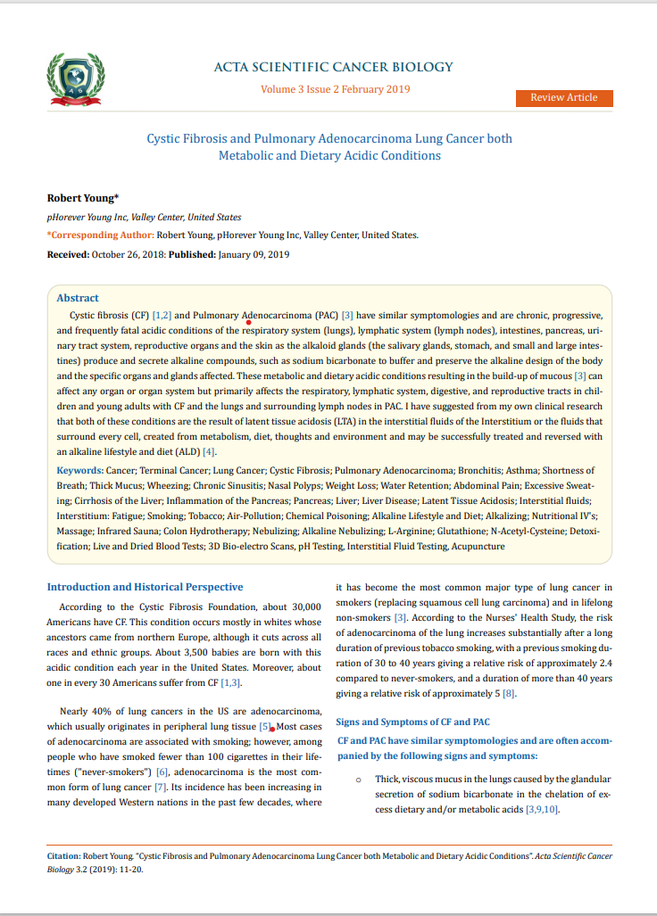

https://www.drrobertyoung.com/post/nano-and-micro-clots-seen-in-the-blood-of-the-vaxxinated-nonvaxxinated
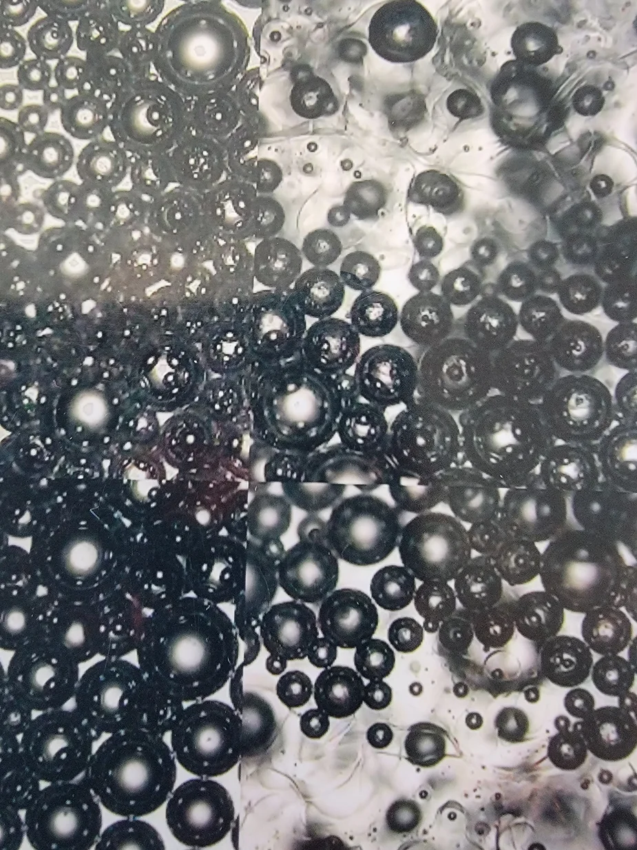
Self-Assembling Graphene Oxide Based Biosensors observed in the unchanged and unstained capillary blood under pHase contrast microscopy of a VAXXinated female.
https://rumble.com/v25lyd2-young-blood-solutions-to-the-pollutions-from-nano-technology.html
References
[1] Asakura, Hidesaku; Ogawa, Haruhiko (2020). “COVID-19-associated coagulopathy and disseminated intravascular coagulation”. International Journal of Hematology. 113 (1): 45–57. doi:10.1007/s12185-020-03029-y. ISSN 0925-5710. PMC 7648664. PMID 33161508.
[2] Khan, Faizan; Tritschler, Tobias; Kahn, Susan R; Rodger, Marc A (2021). “Venous thromboembolism”. The Lancet. doi:10.1016/s0140-6736(20)32658-1. ISSN 0140-6736. PMID 33984268.
[3] Ponti, G; Maccaferri, M; Ruini, C; Tomasi, A; Ozben, T (2020). “Biomarkers associated with COVID-19 disease progression”. Critical Reviews in Clinical Laboratory Sciences. 57 (6): 389–399. doi:10.1080/10408363.2020.1770685. ISSN 1040-8363. PMC 7284147. PMID 32503382.
[4] Velavan, Thirumalaisamy P.; Meyer, Christian G. (25 April 2020). “Mild versus severe COVID-19: laboratory markers”. International Journal of Infectious Diseases. 95: 304–307. doi:10.1016/j.ijid.2020.04.061. PMID 32344011. Retrieved 25 April 2020.
[5] Wells PS, Anderson DR, Rodger M, Forgie M, Kearon C, Dreyer J, et al. (September 2003). “Evaluation of D-dimer in the diagnosis of suspected deep-vein thrombosis”. The New England Journal of Medicine. 349 (13): 1227–35. doi:10.1056/NEJMoa023153. PMID 14507948.
[6] Kogan AE, Mukharyamova KS, Bereznikova AV, Filatov VL, Koshkina EV, Bloshchitsyna MN, Katrukha AG (July 2016). “Monoclonal antibodies with equal specificity to D-dimer and high-molecular-weight fibrin degradation products”. Blood Coagulation & Fibrinolysis. 27 (5): 542–50. doi:10.1097/MBC.0000000000000453. PMC 4935535. PMID 26656897.
[7] Olson JD, Cunningham MT, Higgins RA, Eby CS, Brandt JT (August 2013). “D-dimer: simple test, tough problems”. Archives of Pathology & Laboratory Medicine. 137 (8): 1030–8. doi:10.5858/arpa.2012-0296-CP. PMID 23899057.
[8] Young RO (2016) “Pathological Blood Coagulation and the Mycotoxic Oxidative Stress Test (MOST).” Int J Vaccines Vaccin 2(6): 00048. DOI: 10.15406/ijvv.2016.02.00048
