A UNIVERSAL CURE FOR SEPSIS – AN EPIDEMIC IN DISGUISE!
Where Does All Sickness and Disease Begin?
Answer: In the largest organ of the human body!
What is the largest organ of the human body?
Answer: The Interstitium
Where sepsis sepsis begin?
Answer: The same place cancer, kidney disease, lung disease, Alzheimer’s, lupus, paralysis, diabetes, heart disease, stroke, and all the other 5000 diseases – in the Interstitium that flows through every organ, gland and tissue and touches every human and animal body cell – all 70 trillion!
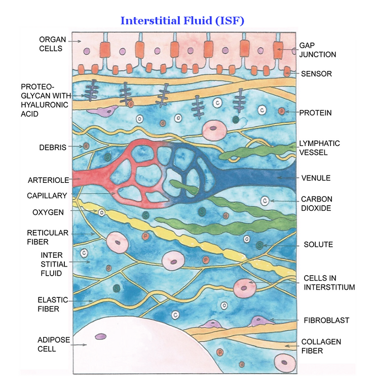
Legend – See below
Organ Cells – The cells at the boundary of the ISF, which supplies the substances to keep it alive.
Gap Junction – It provides cell bonding for multicellular organisms to maintain the body form and structure.
Sensor – This membrane receptor controls the chemical signal and cellular response to and from the cell.
Proteoglycan/Hyaluronic Acid – This large molecule provides viscosity to support the structure.
Protein – This is the nutrient going to the cells.
Acidic Debris – This is the waste draining from the cells.
Lymphatic Vessel – The vessel that carries away some of the debris.
Arteriole – The small branch of the artery carrying oxygenated blood.
Venule – The small branch of the vein carrying de-oxygenated blood.
Capillary – Brings electrons to the cells for energy.
Oxygen – It is released from the capillary to ISF by concentration gradient.
Carbon Dioxide – It becomes HCO3- (bicarbonate, which is a blood pH buffering agent, regulating it within small range of 7.35-7.45 pH – death of the cell at 7.0) in water, reabsorbed into drainages by concentration gradient.
Interstitial Fluid (ISF) – The liquid component of the Interstitium.
Solute – See below
Reticular Fiber – The fibers crosslink to form a fine meshwork to support soft tissues.
Elastic Fiber – Bundle of elastic proteins to prevent deformation.
Fibroblast – Cell that synthesizes the extracellular matrix and collagen.
Adipose Cell – This fat cell – can be derived from undifferentiated fibroblast.
Collagen Fiber – This structural protein is the main component of connective tissue
Cells in the Interstitium – The largest organ of the human body.
Closer examination of the ISF reveals that it contains a mixture of nutrient and acidic waste indicating a primitive origin of the organization. This property is a hallmark of sea water which contains ions of sodium for the transport of electrons to the cells for energy. www.drrobertyoung.com

ABSTRACT
Background:
Every year it is estimated that more than 30 million people worldwide, leading to over 6 million deaths (1) die from potentially preventable systemic acidosis or what is called Sepsis. The burden of systemic acidosis or sepsis is highest in low and middle-income countries where the acid loads from lifestyle and diet on the interstitial fluid compartments of the Interstitium becomes excessive and severe (pH of interstitial fluids drops below 7.2 where normal pH is 7.365). It is estimated that 3 million newborns and 1.2 million children suffer from sepsis globally every year (2).Apr 19, 2018
Why is Sepsis or Systemic Acidosis an Epidemic?
An individual who is sick or injured and not being treated for decompensated acidosis of the Interstitial Fluids of the Interstitium and the blood, buildup excess dietary, metabolic or cellular degenerative acids, in the Interstitium which causes cellular breakdown that leads to Sepsis and then death. Sepsis is suggested in the medical literature to be caused by an outside infection. This so-called infection can only be a contributing factor and not causative. My research, in association with Dr. Galina Migalko, MD, has revealed that Sepsis is an acidic condition of the interstitial fluids of the Interstitium, leading to severe decompensated acidosis and the degeneration of the body cells that make up our organs and glands that sustain life. In addition, acids held in the compartments of the Interstitium can eventually effect the pH of the blood plasma causing erythrocytosis and leukocytosis and eventual death. Therefore, Sepsis is an outfectious condition of the body cells (giving birth to bacteria and/or yeast) and the body’s inability to remove an excessive buildup of acidity in the Interstitium and the blood, through the four channels of elimination, including urination, respiration, defecation or perspiration.
In addition, our research and findings suggest, using high-tech EMF electrode testing of the biochemistry of the body fluids, including the pH of the interstitial fluids of the Interstitium, shows that Sepsis results from an individual’s hyper-inflammatory or hyper-acidic state of the interstitial fluids of the Interstiium and then the blood plasma, created from an acidic lifestyle, diet, metabolism, environment, stress, and injury, all contributing factors.
Currently, Sepsis has a very low Standard of Care in hospitals and hospital-induced outfections that increase the risk of Systemic Sepsis of the Interstitial Fluids of the Interstitium and then the blood plasma.

The cause is directly related to the administration of dextrose, glucose, antibiotic IV’s and acidic hospital food and drink. In response to the low standard of care in hospitals we will share in this article our non-invasive, non-radioactive high-tech medical equipment for testing the chemistry of the blood, interstitial fluids, and intracellular fluids, thereby validating the severe decompensated acidosis, the efficacy of treatment for restoring and balancing the ideal chemistry and alkaline pH at 7.365 and for preventing and treating Systemic Sepsis successfully of the Interstitial Fluids of the Interstitium and Blood Plasma.
Objective:
To examine the biochemistry of the interstitial fluids of the Interstitium, representing 80 percent of the extracellular fluids compared to the biochemistry of the blood plasma, representing 20 percent of the extracellular fluids, to validate that Sepsis is caused by a declining pH of the interstitial fluids leading to decompensated acidosis of the Interstitium and the human blood.

Methods:
Data was drawn from the use of a new technology used by NASA for non-invasive and non-radioactive testing of the chemistry, including the pH, of the blood, interstitial fluids of the Interstitium and the intracellular fluids.

Results:
All patients tested were in hypercalcemia of the interstitial fluids, chronic bone and muscle loss, and decompensated acidosis of the interstitial fluids of the Interstitium with a pH near or below 7.2. After administration of an alkalizing dose of sodium and potassium bicarbonate rectally and/or by IV, the chemistry, including the pH of the interstitial fluids are restored within their normal alkaline range (7.365) resulting in the successful reversal and cure of systemic acidosis or Sepsis, preventing the loss of life.
References
[1] “Sepsis Questions and Answers”. cdc.gov. Centers for Disease Control and Prevention (CDC). 22 May 2014. Archived from the original on 4 December 2014. Retrieved 28 November 2014.
[2] Jui, Jonathan; et al. (American College of Emergency Physicians) (2011). “Ch. 146: Septic Shock”. In Tintinalli, Judith E.; Stapczynski, J. Stephan; Ma, O. John; Cline, David M.; Cydulka, Rita K.; Meckler, Garth D. (eds.). Tintinalli’s Emergency Medicine: A Comprehensive Study Guide (7th ed.). New York: McGraw-Hill. pp. 1003–14. Archived from the original on 15 January 2014. Retrieved 11 December 2012 – via AccessMedicine.
[3] Deutschman CS, Tracey KJ (April 2014). “Sepsis: current dogma and new perspectives”. Immunity. 40 (4): 463–75. doi:10.1016/j.immuni.2014.04.001. PMID 24745331.
[4] Singer M, Deutschman CS, Seymour CW, Shankar-Hari M, Annane D, Bauer M, Bellomo R, Bernard GR, Chiche JD, Coopersmith CM, Hotchkiss RS, Levy MM, Marshall JC, Martin GS, Opal SM, Rubenfeld GD, van der Poll T, Vincent JL, Angus DC (February 2016). “The Third International Consensus Definitions for Sepsis and Septic Shock (Sepsis-3)”. JAMA. 315 (8): 801–10. doi:10.1001/jama.2016.0287. PMC 4968574. PMID 26903338.
[5] Rhodes A, Evans LE, Alhazzani W, Levy MM, Antonelli M, Ferrer R, et al. (March 2017). “Surviving Sepsis Campaign: International Guidelines for Management of Sepsis and Septic Shock: 2016” (PDF). Intensive Care Medicine. 43(3): 304–377. doi:10.1007/s00134-017-4683-6. PMID 28101605.
[6] Jawad I, Lukšić I, Rafnsson SB (June 2012). “Assessing available information on the burden of sepsis: global estimates of incidence, prevalence and mortality”. Journal of Global Health. 2 (1): 010404. doi:10.7189/jogh.01.010404. PMC 3484761. PMID 23198133.
[7] Martin GS (June 2012). “Sepsis, severe sepsis and septic shock: changes in incidence, pathogens and outcomes”. Expert Review of Anti-Infective Therapy. 10(6): 701–6. doi:10.1586/eri.12.50. PMC 3488423. PMID 22734959.
[8] Dellinger RP, Levy MM, Rhodes A, Annane D, Gerlach H, Opal SM, Sevransky JE, Sprung CL, Douglas IS, Jaeschke R, Osborn TM, Nunnally ME, Townsend SR, Reinhart K, Kleinpell RM, Angus DC, Deutschman CS, Machado FR, Rubenfeld GD, Webb SA, Beale RJ, Vincent JL, Moreno R (February 2013). “Surviving sepsis campaign: international guidelines for management of severe sepsis and septic shock: 2012”(PDF). Critical Care Medicine. 41 (2): 580–637. doi:10.1097/CCM.0b013e31827e83af. PMID 23353941. Archived from the original (PDF) on 2 February 2015.
[9] Martin GS, Mannino DM, Eaton S, Moss M (April 2003). “The epidemiology of sepsis in the United States from 1979 through 2000”. The New England Journal of Medicine. 348 (16): 1546–54. doi:10.1056/NEJMoa022139. PMID 12700374.
[10] Poltorak A, Smirnova I, He X, Liu MY, Van Huffel C, McNally O, Birdwell D, Alejos E, Silva M, Du X, Thompson P, Chan EK, Ledesma J, Roe B, Clifton S, Vogel SN, Beutler B (September 1998). “Genetic and physical mapping of the Lps locus: identification of the toll-4 receptor as a candidate gene in the critical region”. Blood Cells, Molecules & Diseases. 24 (3): 340–55. doi:10.1006/bcmd.1998.0201. PMID 10087992.


