Arabic
 Chinese (Simplified)
 Dutch
Dutch English
 French
 German
 Italian
 Portuguese
 Russian
 Spanish
 Swedish
Do YOU Have Filaria Parasites in YOUR Blood & Interstitial Fluids Causing Severe Acute Lung Disease?
Updated: May 15, 2023
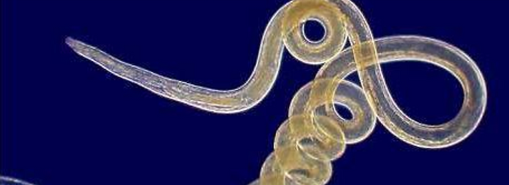
The Filarioidea are a superfamily of highly specialized parasitic nematodes.[1][2] Species within this superfamily are known as filarial worms or filariae (singular “filaria”). Infections with parasitic filarial worms cause disease conditions generically known as filariasis.[3-8]
Currently hundreds of millions of people worldwide, mainly in tropical regions, are infected with pathogenic species of filariae. Where the diseases are endemic many times more are exposed routinely to infection. Some victims harbor more than one medically significant infection simultaneously and this can complicate diagnosis and treatment.[9] This is why I generally recommend a holistic approach using the pH Miracle Lifestyle and Nutrition Protocol to reverse this and other conditions.[4]
Humankind is the definitive host of at least eight species of filariae in various families. Six are particularly significant in medical terms.
The ones that mainly occupy lymph vessels and cause conditions such as adenolymphangitis, elephantiasis, and filarial fever are:
Brugia malayi


Wuchereria bancrofti
Brugia timori
Brugia malaryi

Three other medically important parasitic species are:
Loa loa causes Loa loa filariasis also known as Calabar swelling

Mansonella streptocerca, which causes streptocerciasis, an itchy condition that creates depigmented skin lesions sometimes mistaken for the first signs of leprosy.
Onchocerca volvulus causes cutaneous onchocerciasis and river blindness[9]
The other two which are pathogenic and found in the blood and interstitial fluids of humans.
Mansonella ozzardi

Mansonella perstans

Some Dirofilaria species usually parasitize animals such as dogs, but occasionally infect humans as well. They are not well adapted to humans as hosts and seldom develop properly though they may cause various confusing symptoms.[9]
Various filarial diseases specific to humans are candidates for elimination by such means as breaking the cycle of infection with anti-parasite treatments.[4][11]
The mature worms live in the body fluids and cavities of the definitive hosts, or predominantly in particular tissues. Details vary according to species. Some of the worst pathogens invade vascular, interstitial fluids of the Interstitium and lymphatic vessels and may be numerous enough to clog them.

Some species invade deep connective tissues; some infest subcutaneous connective tissue, causing unbearable itching. Some invade the lungs or serous cavities such as the pleural cavity, pericardial cavity and the interstitial fluids of the lungs. Wherever established, they may survive for years, the fertilized females continuously producing motile embryos called microfilariae rather than eggs.[9][12]

A microfilaria cannot reproduce in the definitive host and cannot infect another definitive host directly, but must make its way through the host’s body to where an intermediate host that acts as a vector can swallow it while itself acting as an ectoparasite to the definitive host. It must succeed in invading its vector organism fairly soon, because, unlike adult filarial worms, microfilariae only survive for a few months to a year or two depending on the species and they develop no further unless they are ingested by a suitable blood-feeding female insect.
In the intermediate host the microfilaria can develop further till the vector conveys it to another definitive host. In the new definitive host the microfilaria complete the final stage of development into sexual maturity; the process takes a few months to a year or more depending on species. The mature filaria then must mate before a female can produce the next generation of microfilariae, so that invasion by a single worm cannot produce an infection. Accordingly, it takes years of exposure to infections before a serious disease condition can develop in the human host.
Once a new generation of microfilariae is released in the primary host, those in turn must seek out host tissue suited to the nature of the vector species. For example, if the vector is a skin-piercing fly such as a mosquito the microfilaria must enter the peripheral blood circulation, whereas species borne by skin-rasping flies such as Simuliidae[12] and skin-cutting flies such as Tabanidae tend to establish in hypodermal tissues. For obscure reasons, some such species actually undergo daily migrations to bodily regions favored by the vector ectoparasites.[5][12] Outside those periods they take refuge in intravascular blood circulation of the lungs.[9]

References
[1] “Filarioidea” (HTML). NCBI taxonomy. Bethesda, MD: National Center for Biotechnology Information. Retrieved 14 January 2019.
[2] Filarioidea at the US National Library of Medicine Medical Subject Headings (MeSH)
[3] “NCBI Taxonomy Browser”. Retrieved 2009-05-21. Filarioidea Taxonomy ID: 6295 Inherited blast name: nematodes Rank: superfamily Genetic code: Translation table 1 (Standard) genetic code: Translation table 5 (Invertebrate Mitochondrial)
[4] Young, RO, Young, SR, “The pH Miracle Revised and Updated (2010). www.phmiracleproducts.com
[5] Janovy, John; Schmidt, Gerald D.; Roberts, Larry S. (1996). Gerald D. Schmidt & Larry S. Roberts’ Foundations of parasitology. Dubuque, Iowa: Wm. C. Brown. ISBN 978-0-697-26071-0.
[6] “Filarioidea”. Animal Diversity Web. Retrieved 14 June 2014.
[7] Wheeler, Lance. “File:Microfilaria of Dirofilaria immitis (Heartworms) Surrounded by Neoplastic Lymphocytes”. Flickr. Retrieved 2 December 2017.
[8] Yoshihito Otsuji. History, Epidemiology and Control of Filariasis. Trop Med Health. 2011 Mar; 39(1 Suppl 2): 3-13. doi:10.2149/tmh.39-1-suppl_2-3 PMC 3153148
[9] Warrell, D. A.; Weatherall, D. J.; Ledingham, J. G. G. (1996). Oxford textbook of medicine. Oxford [Oxfordshire]: Oxford University Press. ISBN 978-0-19-262140-5.
[10] GIDEON Informatics, Inc.; Dr. Stephen Berger (20 January 2017). Mansonelliasis: Global Status (2017 ed.). GIDEON Informatics Inc. pp. 15–. ISBN 978-1-4988-1619-9.
[11] Martial L Ndeffo-Mbah, Alison P Galvani. Global elimination of lymphatic filariasis. The Lancet Infectious Diseases Volume 17, No. 4, p358-359, April 2017 Published: 21 December 2016 DOI: https://dx.doi.org/10.1016/S1473-3099(16)30544-8
[12] O’Donoghue, Peter. PARA-CITE. Published by: School of Molecular & Microbial Sciences, Faculty of Science, The University of Queensland, Brisbane 4072, Australia July, 2010. ISBN 978-1-8649999-1-4 [1]